Medical imaging in clinical trials does far more than confirm a diagnosis. MRI, CT, PET, ultrasound, and X-ray are used to select participants, measure treatment response, understand how a therapy works, and watch for safety signals. Unlike imaging in routine care, trial imaging has to be standardized, reproducible, and defensible, because the images become evidence for regulators. This guide explains how imaging supports each stage of a study, what teams standardize across sites, and how imaging data stays organized from acquisition to endpoint assessment.
How Medical Imaging Is Used Across Clinical Trials
In clinical practice, a radiologist reads a scan to guide care for one patient. In a clinical trial, the same scan is a data point that must be comparable with thousands of others. The National Cancer Institute's Cancer Imaging Program describes an imaging clinical trial as a research study conducted with volunteers, where each study answers specific scientific questions. As Lars Johansson, Chief Scientific Officer at Antaros Medical, puts it, in drug development trials imaging serves to quantify treatment effect in a standardized way that allows for comparisons.
That difference shapes how protocols are written, how images are acquired, and how they are reviewed and archived. Across most trials, imaging plays four distinct roles.

1. Screening Participants and Establishing a Baseline
Before enrollment, imaging often confirms that a participant meets the eligibility criteria in the protocol. In an oncology trial, a CT or MRI may confirm measurable lesions. In a neurology trial, PET may confirm the presence of amyloid plaques. A participant enrolled on the basis of a non-qualifying scan adds variability and can weaken the statistical foundation of the study, so sites need clear acquisition instructions before the first participant is screened.
The baseline scan then becomes the reference point for every later comparison. It must be acquired inside the protocol-specified window before treatment starts, sometimes within days. A late or incomplete baseline leaves the trial without an anchor, which makes it impossible to measure response accurately later. Baseline imaging also documents the participant's disease state at enrollment, which gives essential context if findings change unexpectedly during the study.
2. Measuring Treatment Response and Clinical Trial Endpoints
At each scheduled time point, imaging shows whether the intervention is producing a measurable effect. In oncology, response criteria such as RECIST define exactly how tumors are measured across sequential scans. In other areas, imaging measures plaque reduction, joint damage, or organ perfusion. Each scan has to be acquired under the same conditions as the baseline, because any drift in parameters adds noise that is hard to correct in the analysis.
When an imaging measurement is prespecified as a primary or secondary outcome, it becomes an imaging endpoint. The FDA has published clinical trial imaging endpoint process standards that set out what a defensible imaging workflow looks like, including reader qualification, blinding, and documentation. Endpoints built on that discipline are far easier to defend during regulatory review. For a deeper look at one therapeutic area, see our guide to oncology imaging in clinical trials.
3. Using Imaging Biomarkers to Understand Treatment Effects
Imaging biomarkers are quantifiable features in a scan, such as tumor volume, bone density, lesion count, or tracer uptake, that show how a therapy is acting on the body. They can reveal a biological effect earlier than a clinical outcome would, which gives sponsors mechanistic insight while a trial is still running. Some validated imaging biomarkers are accepted as surrogate endpoints, which can shorten development timelines.
For a biomarker to carry that weight, it has to be validated, measured the same way at every site, and assessed in a blinded and standardized manner. That is why the acquisition protocol, the analysis method, and the reader training matter as much as the biomarker itself. As research published in PMC notes, imaging techniques are increasingly used in oncological clinical trials to provide evidence for decision making, and the same holds in neurology, cardiovascular, and musculoskeletal research.
4. Monitoring Treatment Safety
Imaging also protects participants. Cardiac imaging is routine in trials of agents with cardiotoxic potential, and brain MRI is used in many CNS studies to catch unexpected findings before they become serious adverse events. Safety imaging follows the same protocol discipline as efficacy imaging. A missed time point or a non-compliant acquisition can leave a safety signal undetected, which creates risk for participants and liability for the sponsor and the CRO managing the data.
Taken together, these four roles explain why imaging is planned differently from other trial data. A lab value can be re-run if a sample is retained. A scan cannot be repeated once the participant has moved on to the next treatment cycle, and repeating it exposes participants to additional radiation or burden. Imaging has to be right the first time, which is why so much of the work happens before acquisition rather than after. The choice of modality, the timing of each scan, and the way results will be read should all be decided together, by clinical, imaging, and data management teams working from the same protocol.
Building a Standardized Imaging Workflow Across Trial Sites
Almost every guide to trial imaging says the process must be standardized. Fewer explain what teams actually standardize. In practice, a reliable imaging workflow fixes five things before the first participant is scanned.
- Acquisition parameters. Modality, field strength, sequences, slice thickness, contrast use, and positioning are defined in an imaging manual so that scans from different scanners are comparable.
- Timing. The imaging schedule sets the visit windows for baseline and follow-up scans, and tells sites what to do when a window is missed.
- Site and scanner qualification. Sites demonstrate that their equipment and staff can meet the protocol, often through phantom or test scans, before they scan trial participants.
- Quality control on receipt. Every submitted study is checked for completeness, correct parameters, and image quality, and non-compliant scans trigger a query back to the site.
- Reader procedures. The imaging charter defines who reads the images, how they are trained, what they are blinded to, and how disagreements are resolved.
Standardization pays off because variability does not average out. A different scanner model, a changed protocol, or an inconsistent reading rule can shift a measurement enough to look like a treatment effect or to hide one. Teams that settle these decisions early, ideally while the protocol is still being drafted, avoid the expensive corrections that come from fixing imaging problems after sites are already scanning. Our article on medical imaging strategy covers how to plan for this from the start, and clinical trial imaging compliance explains the regulatory expectations that sit behind these choices.
For a broader view of how imaging fits into modern study operations, download our white paper, Modernizing Image-Driven Clinical Research.

From Medical Images to Reliable Endpoint Assessments
Collecting good images is only half of the job. The other half is turning them into measurements that regulators will accept. That is the purpose of centralized image review, where independent, qualified readers assess images away from the sites that produced them. Central review removes the variability of site-level reads and provides the objective, auditable assessment that many primary imaging endpoints require.
Blinded independent central review (BICR) adds structure to that process. Images are routed to qualified readers in a defined order, readers do not know treatment assignment or site identity, and each measurement and determination is captured in an auditable format. Time-point locking prevents later scans from influencing earlier assessments, and formal adjudication resolves disagreements between primary readers. The result is an endpoint assessment that can be reconstructed step by step during an audit. Our guide to blinded imaging assessments in multicenter studies goes deeper on how these reads are designed, and imaging core labs in clinical trials explains the organizations that usually run them.
Reader consistency deserves its own attention. Even well-trained readers vary, so trials measure that variability directly through intra-reader and inter-reader checks, and they document how discordant reads are adjudicated. Increasingly, AI-assisted tools help with pre-reading, lesion tracking, and quality checks. In a regulated trial, the expectation is that AI supports an expert reader rather than replaces one, and every AI-assisted call remains subject to the same audit trail as a human determination.
Managing Imaging Data Across Multicenter Clinical Trials
Once dozens or hundreds of sites start submitting images, the question changes. It is no longer only whether each scan is good. It is whether the trial can keep every image organized, complete, traceable, and usable from first submission to database lock.
Multicenter trials bring different scanners, software versions, and local habits. Images arrive as DICOM files that must be de-identified consistently, matched to the correct participant and visit, checked against the imaging schedule, and routed to the right readers. Missing time points need to be spotted while a participant can still be scanned, not at lock. Every step, from receipt to query to read to determination, needs a timestamp and an audit trail. As a multicenter guide for clinical research from Collective Minds notes, success depends on standardized imaging parameters, secure data handling, and regular equipment calibration across all participating sites.
Centralizing this work is what makes the resulting dataset trustworthy. When images and metadata live in one governed place, teams can see completeness in real time, resolve queries faster, and reuse the data for secondary analyses after the trial closes. Read more in our article on how to centralize medical imaging data.
Data integrity runs through all of this. Regulators expect imaging records to be attributable, legible, contemporaneous, original, and accurate, and expect systems handling them to be validated and to support electronic records requirements such as 21 CFR Part 11. Access controls, role-based permissions, and complete change histories are not optional extras in a trial that will be inspected. They are part of what makes an imaging dataset acceptable as evidence.
How Clinical Trial Imaging Platforms Support the Workflow
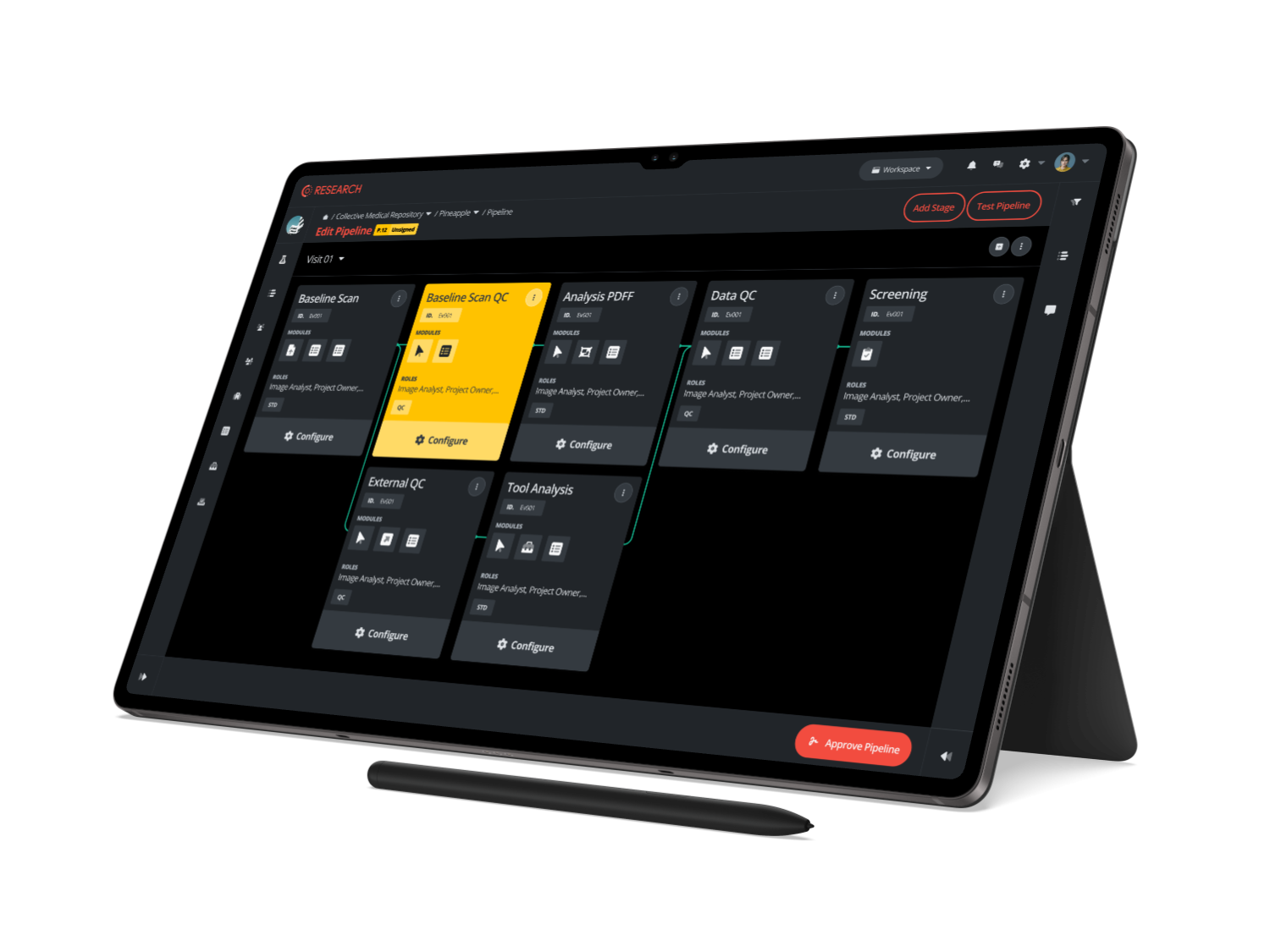
General-purpose file sharing and generic PACS tools were not designed for the demands above. A purpose-built clinical trial imaging platform, often described as an imaging clinical trial management system (ICTMS), brings the workflow into one environment. A well-implemented platform typically provides:
- A centralized, validated image repository accessible to authorized sponsors, sites, and readers
- Automated de-identification and quality control on receipt
- Query management for non-compliant submissions
- Structured central review workflows with blinding, time-point locking, and adjudication
- Complete audit trails for every action on every image
- Integration with EDC and eTMF systems to reduce manual data transfer
Teams that adopt a purpose-built platform early spend less time managing exceptions and more time on the science. If you are comparing options, our overview of imaging vendors for clinical trials outlines the questions worth asking.

Reliable Clinical Trial Imaging Starts Before the First Scan
Imaging in a clinical trial is one of the primary sources of evidence for whether a therapy works, whether a participant is eligible, and whether the study will withstand regulatory review. Every step, from screening through final read, either strengthens that evidence or weakens it.
Teams that treat imaging as an operational afterthought tend to meet the same problems: non-compliant scans, query backlogs, missing time points, and last-minute reconciliation at lock. Teams that plan imaging with the protocol, using standardized acquisition, a centralized platform, and active quality oversight, produce cleaner data with fewer surprises. If you want to see how that looks in practice, talk to the Collective Minds team about your next imaging trial.
Frequently Asked Questions About Medical Imaging in Clinical Trials
What is medical imaging in clinical trials?
Medical imaging in clinical trials is the use of modalities such as MRI, CT, PET, ultrasound, and X-ray within a research protocol to collect standardized, quantifiable data. It is used to screen participants, establish a baseline, measure treatment response, evaluate imaging biomarkers, and monitor safety. Unlike imaging in routine care, the goal is to generate reproducible evidence that can be compared across participants and sites and submitted to regulators.
What imaging modalities are commonly used in clinical trials?
MRI, CT, and PET are the most common, alongside X-ray, ultrasound, and echocardiography. The choice depends on the disease and the endpoint. Oncology trials often rely on CT and MRI with response criteria such as RECIST, neurology trials use MRI and PET for structural and molecular biomarkers, and musculoskeletal trials use X-ray, MRI, and ultrasound to assess joint and tissue changes.
What is an imaging endpoint in a clinical trial?
An imaging endpoint is an outcome measured from medical images and prespecified in the trial protocol, for example change in tumor size, lesion count, or bone erosion score. Imaging endpoints can be primary or secondary. To be accepted by regulators, they need standardized acquisition, validated measurement methods, and blinded, well-documented reading.
What is blinded independent central review in clinical trials?
Blinded independent central review (BICR) is the process in which qualified readers assess trial images without knowing treatment assignment or site identity. It is expected for many primary imaging endpoints because it reduces the bias that can affect site-level reads. BICR workflows include randomized read order, time-point locking, and formal adjudication when primary readers disagree, which produces an objective and auditable assessment.
Reviewed by: Pilar Flores Gastellu on September 30, 2026
.png?width=1024&height=1024&name=Edit%20Pipeline-Galaxy%20Tab%20S8%20Ultra%20(2).png)