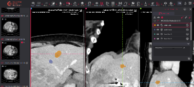
A single CT scan, acquired at a community hospital in the sixth month of a trial, can help decide whether a cancer drug moves forward or quietly dies. On that scan a radiologist measures whether a tumor has grown, shrunk, or held steady, and that measurement, repeated across hundreds of patients and dozens of sites, becomes the evidence a sponsor takes to a regulator. In oncology, imaging is rarely a supporting character. It is often the endpoint itself.
That is also why imaging is one of the most common places an oncology trial springs a leak. A scanner set up slightly differently at one site, a reader applying response criteria a little inconsistently, a timepoint missed because a patient came in a week early, any of these can blur the very signal the trial exists to detect. This guide follows an oncology imaging endpoint from the first design decision to the final analysis, and shows how to build one that holds up. It is written for the clinical operations, imaging, data, and regulatory teams who live with the consequences when it does not.
Key Takeaways
- In many oncology trials the primary or key secondary endpoint is derived from imaging, so the reliability of the imaging directly determines the reliability of the result.
- Building a dependable imaging endpoint is a lifecycle discipline that runs from endpoint definition and modality selection through standardized acquisition, central review, and statistical integration, not a single step near the end.
- The most damaging weaknesses, inconsistent acquisition, reader variability, and missing or off-protocol timepoints, are introduced early and are far cheaper to prevent than to correct.
- A centralized, standardized imaging operation with blinded independent central review is what turns scattered scans into an endpoint a regulator will accept.
Where Imaging Endpoints Influence Oncology Trial Decisions
Imaging shapes oncology trial decisions long before the primary analysis. At screening, scans confirm eligibility and establish the baseline disease that everything else will be measured against. During treatment, follow-up imaging drives the endpoints that define success, from tumor response to disease progression, and it informs the safety decisions that keep patients protected. After the trial, the same images become an asset for exploratory and regulatory work.
The endpoints themselves are familiar to any oncology team. Objective response rate captures how many patients' tumors shrink by a defined amount. Progression-free survival, one of the most consequential imaging-derived endpoints in oncology, measures the time until a tumor grows past a threshold or a new lesion appears. Each of these depends entirely on measurements read off images, which means the quality of those measurements is not a technical detail, it is the endpoint. When a reader marks progression on a scan, that single call can move a patient off a study drug and shift the trial's headline result.
Because so much rides on these reads, oncology imaging clinical trials are held to a high standard of consistency and documentation. A measurement has to mean the same thing whether the scan came from a major academic center or a small regional site, and every step has to be reconstructable later. That is the bar the rest of this guide works toward.
The stakes explain why oncology imaging clinical trials invest so heavily in getting the imaging right. An imaging endpoint that wobbles does not just add noise, it can change the trial's conclusion, delay a submission, or invite questions a regulator will not let go. Treating imaging as core infrastructure, rather than a task delegated to individual sites, is what protects the years of work and investment that sit behind a single readout.
Building Reliable Imaging Endpoints Across the Oncology Trial Lifecycle
A reliable oncology imaging endpoint is built in sequence, and each stage protects the ones that follow. Skip the early decisions and the errors compound, surfacing only when the data is locked and expensive to fix. The stages below trace that sequence from definition to long-term preservation.
Define the Imaging Endpoint Around the Trial Objective
Every imaging endpoint should start from the clinical question the trial is designed to answer. Is the goal to show that tumors shrink, that progression is delayed, or that a functional change occurs before anatomy shifts? The answer determines what is measured, how often, and against which criteria. Defining this precisely up front, in the protocol and the imaging charter, is what keeps every downstream decision aligned.
This is also where the imaging charter earns its place. A well-written charter specifies the endpoint, the timepoints, the modalities, the response criteria, and the review model, so that sites and readers are not left to interpret ambiguous instructions. Vague endpoint definitions are one of the most common root causes of unreliable oncology imaging data, and they are entirely preventable at this stage.
A concrete example clarifies the stakes. If the endpoint is progression-free survival, the charter has to define exactly what counts as progression, how new lesions are handled, and how confirmation scans are scheduled, because a loose definition lets different readers and sites reach different conclusions from the same images. Precision in the charter is what makes the endpoint reproducible in practice.
Select the Appropriate Imaging Modalities and Response Criteria
Oncology imaging is not one method but several, and the disease and endpoint dictate the choice. The response criteria matter just as much as the modality, because they define exactly how an image becomes a measurement. Conventional cytotoxic trials often rely on anatomical assessment, while immunotherapy trials have to account for response patterns, such as pseudoprogression, that older criteria misread. The table below summarizes the frameworks most oncology teams work with.
Choosing the right framework is a clinical and regulatory decision, not a preference. The RECIST 1.1 criteria remain the anatomical backbone of most solid tumor trials, while iRECIST was developed specifically so immunotherapy responses are not misclassified as failures. Getting this choice right early prevents a mismatch between what the trial measures and what the therapy actually does.
Standardize Image Acquisition Across Trial Sites
Once the endpoint and criteria are set, the challenge becomes consistency. A multi-site oncology trial collects images from many scanners, each with its own hardware, software, and local habits. If a lesion is imaged with different slice thickness, contrast timing, or reconstruction settings from one visit or one site to the next, the resulting measurements are not comparable, and the endpoint is quietly undermined.
Standardizing acquisition means specifying and enforcing imaging parameters in the protocol and charter, qualifying sites and scanners before enrollment, and giving sites clear, modality-specific instructions. The goal is that the same patient, scanned at any participating site, produces images a central reader can compare with confidence. This discipline is the foundation of a defensible medical imaging workflow, and it is far easier to build in from the start than to reconstruct after the scans are inconsistent.
Regulators expect this rigor. The FDA's guidance on clinical trial imaging endpoint process standards sets out how sponsors should define acquisition, transfer, and interpretation so that imaging endpoints are credible and reproducible. Aligning the imaging charter with those process standards early removes a common source of regulatory friction later.
Centralize Image Transfer, De-Identification, and Quality Control
Images acquired at sites have to move into a controlled environment where they can be checked before they ever reach a reader. Centralizing image transfer means every scan flows into one system, is de-identified consistently to protect patients, and is checked against the protocol for completeness and quality. A scan that is off-protocol, mislabeled, or of inadequate quality can then be caught and, where possible, repeated while the patient is still available.
This quality control gate is one of the highest-value steps in the entire lifecycle. Catching a problem at intake costs a query, catching it at database lock costs an endpoint. Handling de-identification and quality control on imaging core labs or an equivalent centralized platform is what keeps a distributed trial's data clean enough to analyze.
De-identification in oncology imaging deserves particular care, because tumor tracking depends on preserving the relationships between studies, series, and timepoints even as patient identifiers are removed. Strip too much and a reader cannot follow a lesion across visits, strip too little and patient privacy is at risk. A consistent, automated approach applied uniformly across every site resolves that tension while keeping a complete audit trail.
Design the Central Review and BICR Process
For endpoints that carry regulatory weight, the reads themselves are moved away from the sites to independent, blinded reviewers. Blinded independent central review is the mechanism that removes site-level bias and enforces a single consistent standard across the trial. Readers assess each scan against the predefined criteria without knowing the treatment assignment, and a defined adjudication path resolves disagreements when two readers differ.
Designing this process well means specifying the number of readers, the blinding and randomization of image presentation, the adjudication rule, and the audit trail that records every read. Done properly, blinded independent central review is what lets a sponsor stand behind an imaging endpoint under scrutiny. Done loosely, it becomes the weakest link a regulator will probe first.
The mechanics matter here. Many registrational oncology imaging clinical trials use a two-reader model with a third-reader adjudicator, present images in a randomized, blinded order, and lock the read criteria before the first scan is assessed. These safeguards exist because progression-free survival is especially sensitive to reader bias and timing, and a single inconsistent call can shift the survival curve.
Connect Imaging Results With Clinical and Statistical Data
An imaging read is only useful once it reconciles with the rest of the trial. Response and progression calls have to align with the electronic data capture system, the treatment record, and the statistical analysis plan, so that the imaging-derived endpoint and the clinical dataset tell one coherent story. When imaging data lives in a silo, disconnected from the trial's operational backbone, reconciliation becomes a manual, error-prone scramble at the worst possible time.
Connecting imaging to the trial's systems, including the imaging trial management system and the EDC, is what makes the endpoint analyzable on schedule. It also ensures that when a statistician runs the primary analysis, the imaging data underneath it is complete, traceable, and consistent with everything else.
This connection is also what makes interim analyses and safety oversight possible, since a data monitoring committee can only act on imaging signals that are current, complete, and reconciled with the clinical picture.
Preserve Imaging Data Beyond the Primary Endpoint Analysis
The value of a well-run oncology imaging dataset does not end at the primary readout. Standardized, de-identified, well-documented images support exploratory analyses, imaging biomarker development, AI model training, and future submissions. A dataset that was collected and curated to a high standard becomes a durable asset, while one assembled loosely is difficult to reuse and easy to question.
Preserving this data means retaining it with its full metadata, provenance, and audit trail intact, so that a scan and the decision made on it can be reconstructed years later. This is also where imaging endpoints meet long-term regulatory imaging endpoints requirements, since regulators may revisit the evidence well after the trial closes.
Common Risks That Weaken Oncology Imaging Endpoints
Most failures in oncology imaging endpoints trace back to a small set of preventable risks. Inconsistent acquisition across sites is the most common, and it silently erodes the comparability that measurements depend on. Reader variability is a close second, which is precisely why blinded central review and reader calibration exist. Missing or off-protocol timepoints, often caused by scheduling drift at busy sites, can break a progression-free survival analysis that assumes evenly spaced assessments.
Two more risks deserve attention. Choosing the wrong response criteria, such as applying conventional anatomical criteria to an immunotherapy trial, can misclassify genuine responders as failures and distort the result. And weak data integration, where imaging reads never cleanly reconcile with clinical data, turns the final analysis into a reconciliation project instead of a straightforward readout. The common thread is that all of these are introduced early and cost the most when discovered late. The remedy is the same disciplined, centralized lifecycle described above, applied from the first design decision rather than bolted on at the end.
Strengthen Oncology Imaging Trials With Collective Minds
The story of an oncology imaging endpoint is ultimately a story about control: control over how images are acquired, how they are checked, how they are read, and how they connect to the rest of the trial. Sponsors and CROs that treat imaging as a first-class part of trial design, rather than a service tacked on at the sites, are the ones whose endpoints hold up when it matters.
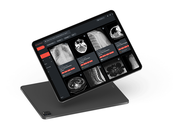
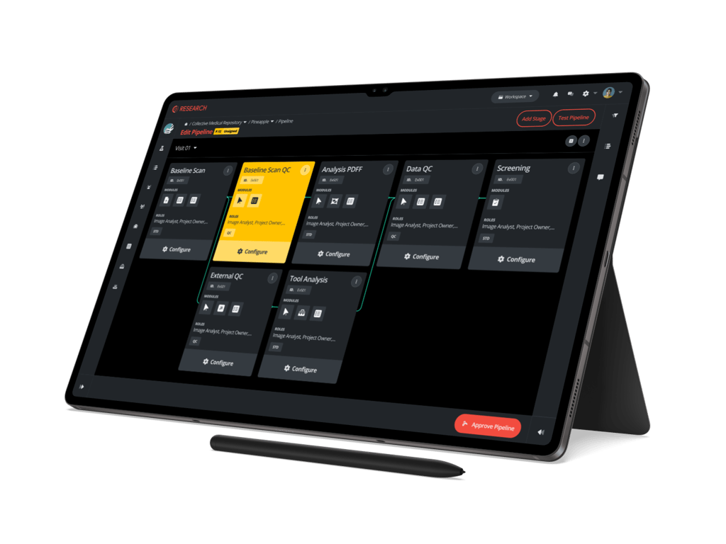
Collective Minds gives oncology teams that control in one place. The platform centralizes image collection from every site, applies consistent de-identification and quality control, supports standardized reads and blinded independent central review, and connects imaging results to the systems that run the trial. Whether you are a sponsor protecting a primary endpoint or an oncology imaging CRO delivering it for a client, the goal is the same: turn scattered scans into imaging endpoints that are reliable, traceable, and ready for regulatory scrutiny. That is how a single CT scan at a small site becomes evidence a whole program can stand on.

Things you might be wondering
What Is the Difference Between an Imaging Endpoint and a Clinical Endpoint?
A clinical endpoint measures how a patient feels, functions, or survives, such as overall survival. An imaging endpoint is derived from measurements read off medical images, such as objective response rate or progression-free survival based on tumor size. Imaging endpoints are often used because they can be assessed earlier and more objectively than some clinical endpoints, but their reliability depends entirely on standardized acquisition and consistent, ideally blinded, central review.
Does Every Oncology Clinical Trial Require BICR?
No. Blinded independent central review is most important for trials where the imaging endpoint carries significant regulatory weight, such as registrational studies using progression-free survival or objective response rate. Early-phase or exploratory studies may rely on local reads or a lighter central review model. The right level of review is a decision made in the imaging charter, based on the endpoint's importance and the level of regulatory scrutiny expected.
Can Artificial Intelligence Be Used to Determine Oncology Imaging Endpoints?
AI is increasingly used to support oncology imaging endpoints, for example by pre-measuring lesions, flagging new findings, or improving consistency between reads. In regulated trials it currently serves as a support to expert readers rather than a replacement, because the endpoint still requires qualified human judgment and a defensible, auditable review process. The strongest workflows combine AI-assisted efficiency with expert central review and clear adjudication.
Reviewed by: Pilar Flores Gastellu on August 10, 2026