Any team training a medical imaging AI model, validating an imaging biomarker, or preparing a research dataset eventually hits the same hard truth: the model is only as good as the labels it learns from. Medical image annotation, the process of marking and describing clinically meaningful features in an image, is what turns raw scans into machine-readable ground truth. Get it right and you have a dataset that supports reliable, defensible results. Get it wrong and no amount of model tuning will recover the errors baked into the labels.
This guide covers medical image annotation as a complete discipline, not just a labeling tool: how it connects clinical expertise to data, how to build a workflow that produces reliable ground truth across modalities, when to bring in medical image annotation services, and how to scale annotation across sites without losing quality or compliance.
Key Takeaways
- Medical image annotation is the bridge between clinical expertise and machine-readable data, and the quality of that annotation sets a hard ceiling on the quality of any downstream AI model or analysis.
- A reliable workflow moves deliberately from clinical objective to source image curation and de-identification, then to method selection, expert calibration, and quality assurance, rather than jumping straight to labeling.
- The right annotation method and workflow depend on both the clinical task and the imaging modality, so a single approach rarely fits every dataset.
- Quality assurance through consensus review and inter-reader agreement is what separates a research-grade dataset from a collection of inconsistent opinions.
- Medical image annotation services and platforms add scale, specialist expertise, and operational capacity when internal teams cannot meet the volume, speed, or compliance demands of a project.
Medical Image Annotation Connects Clinical Expertise With Machine-Readable Data
Medical image annotation is the practice of adding clinically meaningful labels, regions, and measurements to medical images so that they can be analyzed, compared, or used to train and validate algorithms. It is often used interchangeably with labeling, but the two are not identical. Labeling usually refers to assigning a category, while annotation is broader, covering everything from a single classification to a precise pixel-level outline of a lesion. In a clinical context, annotation almost always requires interpretation, which is why it cannot be treated as a purely technical data-entry task.
This distinction has real consequences. A model trained on noisy or inconsistent labels learns those inconsistencies as if they were signal, and the errors are almost impossible to detect after the fact, because there is no cleaner reference to compare against. That is why experienced teams treat annotation quality as a first-class part of model performance, on par with the choice of architecture or the size of the dataset.
The reason annotation matters so much is that it encodes clinical judgment into a form a machine can use. When a radiologist outlines a tumor or grades a finding, they are transferring years of expertise into structured data. That transfer is only trustworthy when it is done consistently and by people qualified to make the call. As the 2024 European Radiology Experimental review by Galbusera and Cina emphasizes, data used to train and validate models should be "consistent, standardised, traceable, correctly annotated, and de-identified." Every one of those properties depends on how the annotation is planned and executed.
For teams working with medical imaging data at scale, this reframes annotation from a downstream chore into a foundational step. The quality of the annotated ground truth determines whether a model generalizes, whether a biomarker holds up under scrutiny, and whether a regulator will accept the evidence. Everything else in the pipeline is built on top of it.
Build a Medical Image Annotation Workflow That Produces Reliable Ground Truth
A dependable annotation workflow is a sequence of deliberate decisions, not a rush to start drawing on images. Skipping the early steps is the most common reason datasets fail validation later. The stages below describe a workflow that consistently produces reliable ground truth.
Define the Clinical Objective and Required Ground Truth
Every annotation project should start with the clinical question, not the tool. What decision will the labeled data ultimately support: detecting a lesion, measuring change over time, classifying disease severity, or something else? The answer determines what counts as ground truth, how precise the annotations must be, and how disagreement between readers will be resolved.
Defining this up front prevents the most expensive kind of rework, which is discovering after thousands of images have been labeled that the labels do not answer the actual research question. A clear objective also tells you the acceptable level of uncertainty, which shapes every later decision about method and review.
Collect, Curate, and De-Identify the Source Images
Before annotation begins, the source images have to be gathered, curated, and cleaned. Curation means selecting images that genuinely represent the clinical problem, removing corrupt or duplicate studies, and confirming that the dataset covers the range of cases the model will encounter. A dataset skewed toward easy cases produces a model that fails on hard ones.
De-identification is not optional. Removing or masking protected health information, from DICOM metadata to identifiers burned into the pixels, protects participants and satisfies privacy obligations. Robust DICOM anonymization has to be done consistently across the whole dataset while preserving the information the project depends on, such as study, series, and timepoint relationships. Done properly, it also underpins imaging compliance for any work that will face regulatory review.
Select the Right Annotation Method for the Clinical Task
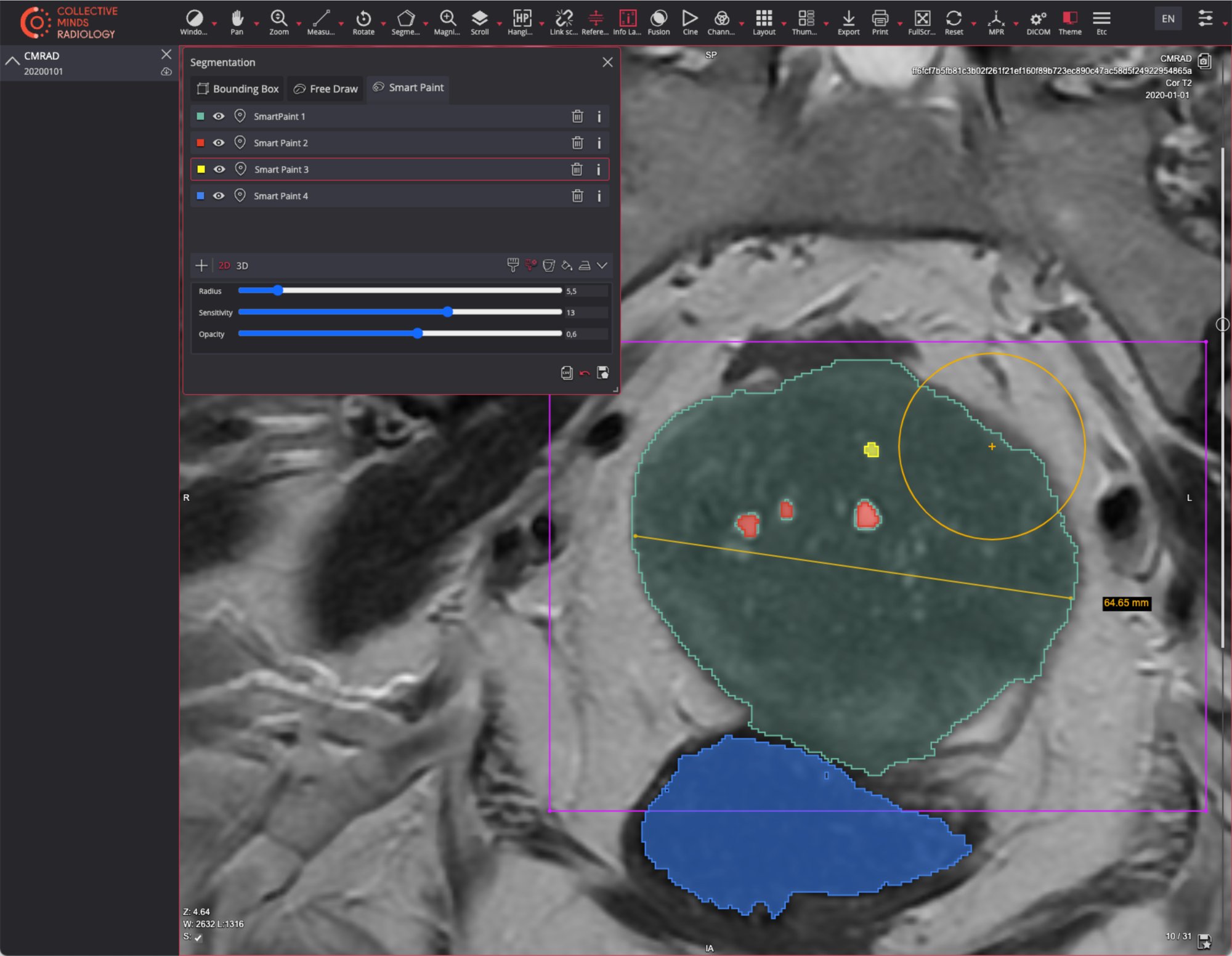
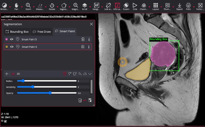
Annotation is not one technique but a family of them, and the clinical task dictates the choice. Image-level classification assigns a single label to a whole study, such as normal versus abnormal. Bounding boxes localize a finding without describing its exact shape. Segmentation, whether a rough region or a precise pixel-level mask, captures the true boundary of a structure or lesion and is essential for volume and change measurement. Landmarks mark specific anatomical points, and measurements capture distances, diameters, or standardized scores.
Each method carries a different cost in time and expertise, and a different level of information. Pixel-level segmentation is far more informative than a bounding box but also far slower to produce and harder to keep consistent. Matching the method to the objective, rather than defaulting to the most detailed option, keeps a project both rigorous and feasible.
A concrete example makes the tradeoff clear. In an oncology study that tracks tumor burden, a diameter measurement following a standard such as RECIST may be enough to support the endpoint, and it is fast and reproducible. If the goal is instead to train a model to segment tumors automatically, the same images need full volumetric masks, which take far longer to produce and demand tighter quality control. The clinical objective, not habit, should decide which one you invest in.
Adapt the Workflow to Each Imaging Modality
A workflow that works for chest X-rays will not transfer unchanged to volumetric CT, MRI, PET, ultrasound, or digital pathology. Cross-sectional modalities like CT and MRI produce three-dimensional volumes, so a lesion has to be annotated across many slices, and readers need tools that maintain spatial consistency through the stack. Functional and multi-parametric studies add channels of information that annotators must interpret together.
Digital pathology and whole-slide imaging bring gigapixel images and their own file conventions, while ultrasound introduces operator-dependent variability that annotation guidelines have to account for. The practical implication is that modality-specific considerations, including resolution, dimensionality, and file format, need to be settled during workflow design, not improvised mid-project.
Nuclear medicine and PET add a further layer, because meaningful annotation often depends on quantitative uptake values rather than anatomy alone, and readers may need co-registered anatomical images for reference. The broader point is that dimensionality, resolution, dynamic range, and file conventions all vary by modality, and a workflow that ignores those differences will produce inconsistent labels that only surface as problems during model validation.
Create Annotation Guidelines and Calibrate Expert Readers
Consistency is impossible without a written guideline that defines exactly what to annotate, how to handle ambiguous cases, and what to do at the edges of a finding. The guideline is the single source of truth that keeps multiple readers aligned. Without it, each annotator quietly applies their own interpretation, and the resulting dataset carries hidden contradictions.
Calibration turns the guideline into practice. Before full-scale annotation, readers work through a shared set of training cases, compare results, discuss disagreements, and refine the guideline until they converge. This upfront investment dramatically reduces variability later and is especially important when annotators are distributed across institutions or when the reader pool changes over the life of a project.
Annotate Images Within Their Native Clinical Context
Annotations are most reliable when readers see the image the way they would in clinical practice, with the correct windowing, the surrounding slices, relevant priors, and, where appropriate, clinical context. Stripping an image down to a flat picture for a generic labeling tool discards exactly the information an expert relies on to make an accurate call.
Annotating in a proper medical imaging environment also preserves the link between the annotation and the original study, its metadata, and its provenance. That traceability is what lets a team reconstruct, months later, exactly how a given label was produced, which is indispensable for both quality audits and regulatory defensibility.
Validate Annotations Through Quality Assurance and Consensus Review
No annotation is complete until it has been checked. Quality assurance can combine automated checks for obvious errors, such as empty or out-of-range labels, with expert review of a sample or the whole set. For tasks where interpretation varies, having multiple readers annotate the same images and measuring their agreement, using metrics like the Dice coefficient for overlap or kappa for categorical agreement, quantifies reliability rather than assuming it.
Where readers disagree, a defined adjudication path, often a senior reader or a consensus panel, resolves the case and feeds the lesson back into the guideline. This consensus process, borrowed directly from how central reads are run in clinical trials, is what elevates a dataset from a set of individual opinions to a defensible ground truth. It is the difference between medical image data annotation that survives scrutiny and annotation that quietly undermines every result built on it.

Medical Image Annotation Services Add Scale, Expertise, and Operational Capacity
Even a well-designed workflow can outstrip an internal team's capacity. When a project needs tens of thousands of annotated studies, rare-subspecialty expertise, fast turnaround, or documented compliance that an internal group cannot easily provide, medical image annotation services and managed platforms become the practical answer. The decision is rarely all or nothing, most teams blend internal control over clinically critical labels with external capacity for volume.
The economics reinforce that hybrid approach. Building and retaining an internal pool of qualified readers is expensive and slow to scale, while relying entirely on an external vendor can distance a team from the clinical nuances of its own protocol. Many organizations therefore keep the definition of ground truth, the guidelines, and final adjudication in house, while outsourcing the high-volume, well-specified annotation that follows. This preserves clinical control where it matters and buys scale where it does not.
The table below compares the two approaches across the dimensions that usually drive the decision.
When evaluating an external partner, the questions that matter most are about rigor, not just price: How are annotators qualified and calibrated? What quality assurance and consensus process is in place? How is de-identification handled, and can every annotation be traced back to its source? A provider that cannot answer these clearly is a risk to the entire dataset.
Scale Medical Image Annotation Across Sites With Collective Minds
Scaling annotation across multiple sites is where many projects struggle, because consistency, security, and traceability all get harder as more people and locations are involved. This is precisely the problem a purpose-built medical imaging platform is designed to solve. Rather than exporting flat images into a generic labeling tool, teams annotate within a controlled environment that preserves the full clinical study, its metadata, and its audit trail.
Collective Minds provides standardized annotation workflows, consensus and reader management, and de-identification within a single platform, so that centralized imaging data stays consistent and compliant no matter how many sites contribute. For sponsors and research teams that also rely on expert reads, the same infrastructure supports the kind of controlled, documented review that imaging core labs provide, connecting annotation directly to the clinical and regulatory standards a trial has to meet.
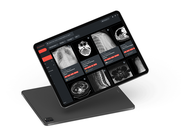
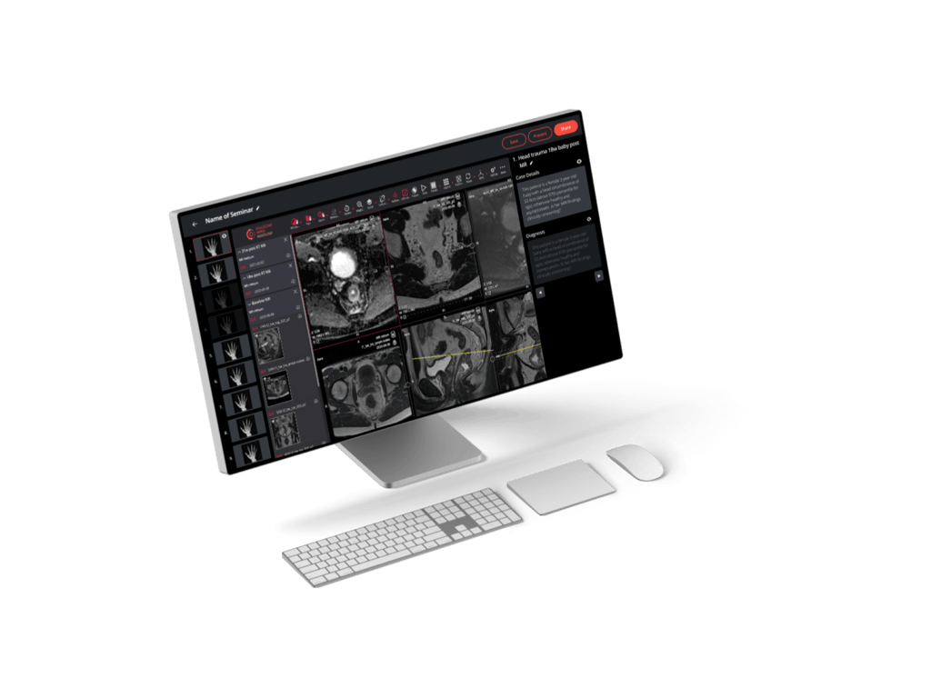
Medical image annotation, segmentation, and labeling in Collective Minds Research.
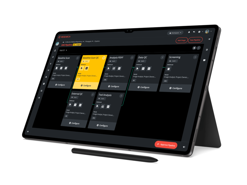
Standardized annotation and labeling workflows in Collective Minds Research.
Build Annotation Around the Clinical Decision, Not the Labeling Tool
Medical image annotation is often treated as a tooling problem, as if the right software were enough to guarantee a good dataset. The evidence points the other way. Reliable ground truth comes from a disciplined workflow: a clear clinical objective, carefully curated and de-identified images, the right method for the task and modality, calibrated expert readers, annotation in native clinical context, and rigorous quality assurance. The tool matters only insofar as it supports that discipline.
For teams building AI-ready datasets or imaging endpoints, the practical takeaway is to design the annotation process around the clinical decision it ultimately serves, and to choose infrastructure and partners that reinforce consistency, traceability, and compliance at every step. That is what turns a pile of images into evidence you can trust and defend.

Things you might be wondering
What Qualifications Should Medical Image Annotators Have?
It depends on the task, but clinically meaningful annotation generally requires readers with relevant medical or imaging expertise, such as radiologists, pathologists, or trained specialists, especially for interpretation-heavy work like lesion segmentation or disease grading. Simpler tasks can involve trained non-experts working under expert-defined guidelines and review. What matters most is that annotators are calibrated against a shared guideline and that their work is quality-checked.
Can Artificial Intelligence Automate Medical Image Annotation?
Partly. AI-assisted annotation can accelerate the work by pre-segmenting structures or suggesting labels that a human then reviews and corrects, which is often faster than annotating from scratch. It does not remove the need for expert oversight, because automated labels still carry errors and require validation. In practice, the strongest workflows combine automation for speed with expert review and consensus for reliability.
How Long Does Medical Image Annotation Take?
There is no single answer, because time scales with the method, the modality, and the quality bar. Image-level classification can take seconds per study, while precise pixel-level segmentation of a volumetric scan across many slices can take much longer, and consensus review adds further time. Planning realistic timelines means accounting for curation, guideline creation, calibration, and quality assurance, not just the labeling itself.
Which File Formats Are Used for Medical Image Annotation?
DICOM is the dominant standard for medical images, and annotations are frequently stored in DICOM-native objects such as segmentation and structured report objects, which keep the annotation linked to the original study. Other formats, including NIfTI, are common in research and AI pipelines, and pathology uses whole-slide formats. Preserving the connection between the annotation and the source study, whatever the format, is essential for traceability.
Reviewed by: Pilar Flores Gastellu on September 9, 2026