
A scan comes off a scanner in Warsaw at nine in the morning. By the time the coordinating site in Boston opens for the day, a qualified reader needs to have already looked at it. That handoff is not a scientific problem. It is a logistics one: getting an image from one country to a reader in another, on time, without any loss of image quality along the way. The trial's endpoint is built on that image, so if quality slips in transit, so does the measurement the whole study depends on. In a study built around cardiac safety or infection markers, a slow handoff is a minor inconvenience. In a neuroscience trial, where the imaging read is often the endpoint itself, it can decide whether a patient is randomized on schedule and whether the data package still holds up a year later.
Brain imaging has moved from a supporting measurement to the backbone of how central nervous system, or CNS, drug development proves that a therapy does what it claims. Volumetric MRI tracks whether a neurodegenerative disease is progressing. PET imaging shows whether a drug is reaching its target in the brain. Structural and functional scans screen patients into the right trial arm and flag safety signals before they become adverse events. None of that works without an imaging workflow built for the job.
This guide walks through how brain imaging supports CNS clinical trials: the biomarkers and modalities in use today, the operational realities of running imaging across dozens of sites and several trial phases, and what a sound imaging strategy looks like for a CNS program.
How Brain Imaging Is Used Across CNS Clinical Trials
Neuroimaging shows up at nearly every stage of a CNS trial, from screening a candidate at baseline to confirming a treatment effect at the final visit. Sponsors and CROs increasingly treat imaging in CNS clinical trials as a core part of the evidence package rather than a secondary measurement layered on top of clinical rating scales. The value it adds breaks down into a few distinct jobs, each with its own imaging and workflow requirements.
Using Imaging Biomarkers to Measure Disease Biology and Treatment Effects
Quantitative imaging biomarkers let a sponsor measure disease biology directly instead of relying only on clinical rating scales, which are subjective and can vary between raters and visits. Brain volume and atrophy on structural MRI track neurodegeneration over time. Amyloid and tau PET quantify the protein pathology behind Alzheimer's disease, an approach that public research initiatives such as the Alzheimer's Disease Neuroimaging Initiative helped establish as a standard part of how the field measures disease progression. Dopamine transporter imaging, cleared by the FDA for differentiating parkinsonian syndromes, helps characterize dopaminergic loss in Parkinson's programs. These measurements increasingly serve as secondary or exploratory endpoints, and in specific indications, regulators have accepted a strong imaging biomarker result as a surrogate endpoint reasonably likely to predict clinical benefit, which is the basis several recent Alzheimer's therapies used for accelerated approval.
The appeal of an imaging biomarker is that it measures something physical rather than something reported. A clinical rating scale depends on a rater's judgment on a given day, which introduces variability that has nothing to do with the drug being tested. A volumetric MRI measurement or a PET signal reflects tissue and biology directly, which is part of why regulators have grown more comfortable accepting imaging endpoints in CNS programs where a reliable clinical measure is hard to standardize across sites and raters.
Supporting Target Engagement and Patient Selection in CNS Trials
PET-based target engagement studies confirm that a drug is actually reaching and binding its intended receptor or protein in the brain, which informs dose selection early in development rather than after a Phase II readout falls flat. Imaging also drives enrollment criteria. Amyloid-PET positivity, for example, is used as a gate in Alzheimer's trials to enroll patients who actually carry the pathology a drug targets. Getting that gate right matters. Enrolling the wrong population is one of the more expensive and preventable ways a CNS trial fails.
The cost of getting patient selection wrong shows up months or years later, when a trial reaches its primary analysis and the effect size is diluted by patients who never had the underlying pathology to begin with. An imaging-based screening gate front-loads that risk into the enrollment process, where it is far cheaper to manage. It also protects patients from participating in a study that was never going to help them, which is its own reason to get the imaging criteria right at the design stage rather than treating them as a formality.
Monitoring Disease Progression and Treatment Safety With Imaging
Serial MRI tracks how a disease progresses over the course of a trial, giving sponsors a biological trajectory to set against the clinical one. Imaging also carries real safety responsibilities. Amyloid-related imaging abnormalities, a known risk with amyloid-targeting therapies, require frequent, standardized MRI reads on a schedule tight enough to catch a finding before it becomes a safety event rather than after. That is only possible when imaging moves fast enough for a read to happen while the finding, and the patient, are still actionable.
Progression monitoring and safety monitoring often rely on the same scan, read for two different purposes. A routine surveillance MRI in an amyloid-targeting trial is checked for signs of ARIA and, at the same time, feeds the longitudinal volumetric measurement the trial uses to track disease course. Splitting those reads across disconnected systems or vendors adds a step that safety monitoring, in particular, cannot afford to lose time to.
Applying MRI, PET, SPECT, and Other Imaging Modalities in Clinical Trials
Modality choice depends on the biological question. Structural MRI measures anatomy and volume. Functional MRI maps brain activity and connectivity. PET quantifies molecular targets, from amyloid and tau to receptor occupancy. SPECT, including dopamine transporter imaging, supports movement disorder trials where PET access is limited. Diffusion imaging characterizes white matter integrity in conditions where connectivity itself is part of the disease. None of these modalities are interchangeable, and a CNS program often uses more than one across its lifecycle. What ties them together operationally is the need to standardize acquisition parameters across scanners, field strengths, and sites so that a measurement taken in one country means the same thing as the same measurement taken in another. A 3-tesla scanner and a 1.5-tesla scanner do not produce identical volumetric measurements without correction, and a PET tracer protocol that varies between sites can shift a quantitative result enough to blur a real treatment effect. Getting the modality right is a scientific decision. Keeping it consistent once the trial is running is an operational one, and it is usually the harder of the two.
Using Brain Imaging Across Different Clinical Trial Phases
Imaging requirements scale with the phase of the program. Phase I trials lean on imaging for safety monitoring and early target engagement or pharmacokinetic and pharmacodynamic data. Phase II trials use biomarker change as proof of concept before committing to a larger, more expensive study. Phase III trials need registration-grade rigor: standardized protocols, qualified readers, and a review process that can withstand regulatory scrutiny. Phase IV work shifts toward long-term safety monitoring in a broader, less controlled population. A platform or vendor that works well for an early, single-site Phase I study will not automatically hold up to a 40-site Phase III program, which is why imaging needs get reassessed at every phase transition rather than assumed to carry over.
That reassessment usually touches reader capacity as much as technology. A Phase I study can run on a small pool of specialist readers. A global Phase III program needs that capacity to scale by an order of magnitude, across time zones, without the review queue backing up. Planning for that jump early, rather than scrambling to add reader capacity mid-study, is one of the more common gaps between a CNS program's imaging plan on paper and how it actually performs once enrollment ramps up.

Managing Imaging Data in Multi-Center CNS Clinical Trials
Multi-center CNS trials add a layer of difficulty that a single-site study never has to solve. Different sites run different scanner makes, different field strengths, and, left unmanaged, different acquisition protocols. A biomarker that is precise within one site can become noisy the moment it is pooled across several, not because the biology changed but because the images were never comparable to begin with. Getting multi-center imaging trials right means standardizing acquisition protocols before the first patient is scanned, running phantom calibration across sites, and centralizing quality control so a deviation is caught at the site that produced it rather than discovered months later during analysis.
The workflow around the data matters as much as the imaging science. Manual transfer between sites, sponsors, and reading centers introduces delay and version confusion at exactly the moments when a fast, clean handoff matters most: screening decisions, safety reviews, and interim analyses. A centralized flow, where images move from the scanner into a controlled platform without manual routing, keeps a multi-site dataset consistent enough to support the statistical power the trial was designed around.
Site turnover adds another layer most CNS programs underestimate. A three-year study can see several changes in site staff, project managers, or even imaging equipment over its lifetime. Every one of those transitions is a chance for a protocol deviation to slip through unnoticed, unless the imaging workflow itself, rather than the memory of whoever set it up, is what enforces the standard. Centralizing acquisition checks and quality control at the platform level means a new site coordinator inherits a workflow that already knows what a compliant scan looks like, instead of relying on documentation that may or may not have been read.
Building an Effective Imaging Strategy for CNS Drug Development
A sound medical imaging strategy for a CNS program starts well before the first site is activated. The endpoint and the biomarker need to be defined together, with imaging specialists involved in that conversation rather than brought in after the protocol is locked. Modality and acquisition protocol have to be standardized across every site that will contribute data, with the calibration and training to back it up. Central review needs qualified neuroradiologists or imaging experts reading against a consistent, blinded standard, not a patchwork of site-level interpretations.
Two other pieces round out the strategy. Imaging has to integrate cleanly with the rest of the trial's systems, so a biomarker result reconciles with the clinical data instead of living in a separate silo. And every step, from acquisition to the final reported endpoint, needs a validated, traceable audit trail. Meeting clinical trial imaging compliance requirements is not a separate task bolted onto the imaging plan. It is what makes the imaging plan usable when the study reaches submission.
The teams that get this right tend to treat imaging strategy as a discipline of its own, sitting alongside biostatistics and clinical operations rather than reporting up through either one. That has a practical effect on timelines. Decisions about modality, reader qualification, and data flow get made once, early, with the people who understand the tradeoffs, instead of getting revisited every time a new site raises a question the protocol didn't anticipate.

Advancing CNS Clinical Trials With Better Imaging Workflows
Most of the friction in CNS imaging trials does not come from the science. It comes from what surrounds it: images moved manually between sites and sponsors, quality checks that happen after a patient has already left the site, systems that do not talk to each other, and a new project manager every six months on a three-year study. None of that friction improves quality. It adds delay, adds risk, and reduces visibility into a study that is already hard to run.
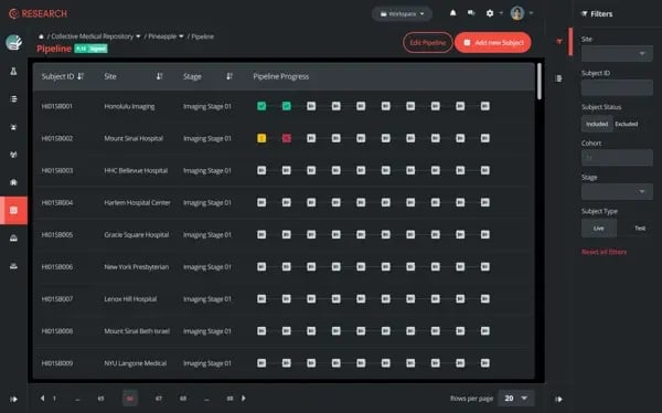
The fix is not more process. It is removing what slows a trial down while keeping everything that protects its rigor. Collective Minds Research connects scanners directly to a centralized platform, so imaging data flows automatically from acquisition into the research pipeline instead of waiting on manual handoffs. A pre-connected network of 300+ imaging sites and access to 30,000+ imaging experts across 30+ countries means CNS programs get qualified neuroradiology and specialist reads without building that capacity in-house. Imaging core labs and specialist reviewers can be brought into a study through the same network, rather than sourced separately for every trial. And automated medical imaging workflows keep the audit trail intact from the scanner to the reported endpoint, so speed and compliance move together instead of trading off against each other.
Less friction, stronger evidence. That is what a CNS program needs from its imaging workflow, and it is the standard worth building toward for any trial where the brain scan is the evidence the outcome depends on.

Things you might be wondering
How are imaging endpoints selected for CNS clinical trials?
Imaging endpoints are selected based on the biological question the trial needs to answer and the modality best suited to measure it. A trial studying neurodegeneration might use volumetric MRI to track brain atrophy, while an amyloid-targeting therapy trial relies on amyloid PET. The choice is made early, in collaboration with imaging specialists and biostatisticians, and needs to align with regulatory expectations for the specific indication before the protocol is finalized.
What challenges affect the use of imaging in neuroscience clinical trials?
The most common challenges are variability across scanners and sites, slow and manual data transfer, inconsistent acquisition protocols, and reader variability in interpreting complex neuroimaging findings. Multi-center CNS trials are especially exposed to these issues because a biomarker has to remain comparable across every site contributing data, not just accurate within any single one.
How can clinical trial teams improve imaging data consistency across multiple sites?
Consistency starts with standardized acquisition protocols and phantom calibration across every scanner in the trial, backed by site training before the first patient is scanned. Centralizing quality control and review, rather than leaving interpretation to individual sites, catches deviations early and keeps the dataset comparable when it is pooled for analysis.
When should imaging specialists be involved in CNS trial planning?
Imaging specialists should be involved while the protocol is still being drafted, not after it is locked. Decisions about which biomarker to use, which modality fits the biological question, and how acquisition will be standardized across sites are far cheaper to get right at the design stage than to correct once sites are already scanning patients.
How does imaging data support research beyond the original clinical trial?
Imaging datasets collected under a standardized, well-documented protocol often have value beyond the trial they were built for. Properly annotated and stored imaging can support secondary analyses, natural history research, and future biomarker validation work, provided the original acquisition and quality control were rigorous enough to make the data trustworthy outside its original context.
Reviewed by: Mathias Engström and Hilde Andersen on August 26 2026